File:Enamel looks like wood.tif

File originale (1 920 × 1 200 pixel, dimensione del file: 6,72 MB, tipo MIME: image/tiff)
| Questo file e la sua pagina di descrizione (discussione · modifica) si trovano su Wikimedia Commons (?) |
Dettagli
| Descrizione |
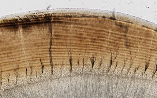
English: This image contains most Enamel's main structures.
The long and curved brown section consists of enamel prisms and it is possible to see the array they present. Enamel prisms are made up of hidroxiapatite. At the lower part of enamel, we can appreciate enamel tufts (the grassy and branchy structures). Enamel tufts run in the same direction as enamel prisms and their grassy appearance is due to the fact that they are hipomineralized structures. Almost in the middle of the picture, a straight line goes from bottom to top of the enamel. This is a lamellae, lamellae's cross enamel in all of its thickness. It is also possible to see the enamel-dentine junction that sits right between the enamel (brown part) and the dentine (grey and finely striped section underneath). This junction keeps both structures together and its easy to spot since it looks like a bumpy area between the the enamel and the dentine. To capture this image with the highest detail possible, a Software tool was used (Extended focal imaging in Cell Sens Software) this tool allows to create a single in-focus image from successive image planes as the focus knob is turned. This image was taken using Brightfield Microscopy and a 40x objective. |
| Data | |
| Fonte | Opera propria |
| Autore | Etenia Urquiza |
Licenza
- Tu sei libero:
- di condividere – di copiare, distribuire e trasmettere quest'opera
- di modificare – di adattare l'opera
- Alle seguenti condizioni:
- attribuzione – Devi fornire i crediti appropriati, un collegamento alla licenza e indicare se sono state apportate modifiche. Puoi farlo in qualsiasi modo ragionevole, ma non in alcun modo che suggerisca che il licenziante approvi te o il tuo uso.
- condividi allo stesso modo – Se remixi, trasformi o sviluppi il materiale, devi distribuire i tuoi contributi in base alla stessa licenza o compatibile all'originale.
Cronologia del file
Fare clic su un gruppo data/ora per vedere il file come si presentava nel momento indicato.
| Data/Ora | Miniatura | Dimensioni | Utente | Commento | |
|---|---|---|---|---|---|
| attuale | 22:26, 14 dic 2019 | 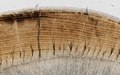 | 1 920 × 1 200 (6,72 MB) | Etenia Urquiza | User created page with UploadWizard |
Pagine che usano questo file
La seguente pagina usa questo file: